Like every other week, the first thing I noticed was that most of the water in my aquarium had evaporated. Also, a lot of my organisms from the first week have completely died off. There are forgotten skeletons of rotifers and cyclopses near and throughout the bit of sediment on the baseline of the aquarium. These organisms have now been replaced by diatoms. There are literally diatoms EVERYWHERE!! Today I also was able to identify an Arcella Artocrea. An Arcella is typically enclosed in a chitinous, umbrella-shaped test (or shell) that has a single central aperture through which the pseudopods – which are used for locomotion – extend out. In some species the aperture is surrounded by a ring of pores (Wiki). There were also a few rotifers still active in the open areas of the aquarium.
Botany 111: An Inquiry Into The Dynamic Microorganisms In Our Environment
General Botany 111, Section 001
Thursday, November 10, 2011
Thursday, November 10, 2011
This week concluded our observations for our microaquariums. Although this is true, I still think I will continue to observe my aquarium; at least until after Thanksgiving break.
Like every other week, the first thing I noticed was that most of the water in my aquarium had evaporated. Also, a lot of my organisms from the first week have completely died off. There are forgotten skeletons of rotifers and cyclopses near and throughout the bit of sediment on the baseline of the aquarium. These organisms have now been replaced by diatoms. There are literally diatoms EVERYWHERE!! Today I also was able to identify an Arcella Artocrea. An Arcella is typically enclosed in a chitinous, umbrella-shaped test (or shell) that has a single central aperture through which the pseudopods – which are used for locomotion – extend out. In some species the aperture is surrounded by a ring of pores (Wiki). There were also a few rotifers still active in the open areas of the aquarium.
Like every other week, the first thing I noticed was that most of the water in my aquarium had evaporated. Also, a lot of my organisms from the first week have completely died off. There are forgotten skeletons of rotifers and cyclopses near and throughout the bit of sediment on the baseline of the aquarium. These organisms have now been replaced by diatoms. There are literally diatoms EVERYWHERE!! Today I also was able to identify an Arcella Artocrea. An Arcella is typically enclosed in a chitinous, umbrella-shaped test (or shell) that has a single central aperture through which the pseudopods – which are used for locomotion – extend out. In some species the aperture is surrounded by a ring of pores (Wiki). There were also a few rotifers still active in the open areas of the aquarium.
Thursday, November 3, 2011
Thursday, November 3 Observation
On Thursday, November 3rd, I observed my microaquarium for the third time. This week i was a bit disappointed to find out that many of my organisms had died. There were many empty shells/skeletons of once living cyclopses and rotifers on the bottom layer of my aquarium; mixed in with the thin layer of soil. Although many of my organisms seemed to either be hiding or no longer living, i was still able to identify a few new organisms.
-I identified a Litonotus
-I also identified with Dr. McFarland's help, two Euchlanis (a type of rotifer)
-I also was able to get a picture of a large group of Diatoms. The Diatoms seem to be taking over all the once open space in my aquarium
-I identified a Litonotus
-I also identified with Dr. McFarland's help, two Euchlanis (a type of rotifer)
-I also was able to get a picture of a large group of Diatoms. The Diatoms seem to be taking over all the once open space in my aquarium
Friday, October 28, 2011
Information On The Food Pellet
On Friday October 21, 2011 "ONE" Beta Food Pellet was inserted into each microaquarium.
On your blog posting for this week include date the food pellet was added along with the following information: "Atison's Betta Food" made by Ocean Nutrition, Aqua Pet Americas, 3528 West 500 South, Salt Lake City, UT 84104. Ingredients: Fish meal, wheat flower, soy meal, krill meal, minerals, vitamins and preservatives. Analysis: Crude Protein 36%; Crude fat 4.5%; Crude Fiber 3.5%; Moisture 8% and Ash 15%.
-Ken McFarland
Wednesday, October 26, 2011
Monday, October 24th Observation
On Monday, October 24th I observed my micro-aquarium for a second time. The first thing i noticed upon entering the lab was that some of the water in the aquarium had evaporated. Dr. McFarland said that we would always have to add more water after observing the aquariums. I also noticed that there was something circular suspended in the water. Dr. McFarland also informed me that it was a data food pellet that had been added to our aquariums over the past weekend.
Information on the Food Pellet
-"Atison's Betta Food" made by Ocean Nutrition, Aqua Pet Americas, 3528 West 500 South, Salt Lake City, UT 84104.
-Ingredients: Fish meal, wheat flower, soy meal, krill meal, minerals, vitamins and preservatives.
-Analysis: Crude Protein 36%; Crude fat 4.5%; Crude Fiber 3.5%; Moisture 8% and Ash 15%.
Findings under the microscope:
-There were many more organisms in my micro-aquarium then last week.
-With Dr. McFarland's help, I identified a flat worm, flagellates, more rotifers, a huge group of diatoms, a seed shrimp, cellias, and a cyclops!
-I was able to get a picture of all of them as well.
Definitions:
-Flagellate: Flagellates are single-celled protists with one or more flagella, whip-like organelles often used for propulsion. The flagella is used for movement through the liquid. Some flagellates live as colonial entities, while others function as a single cell. Most are free-living organisms, however, a number are parasitic or pathogenic for animals and humans. They multiply by binary fission and some species posses cyst stages.
-Rotifer: Rotifers are multicellular animals with body cavities that are partially lined by mesoderm. These organisms have specialized organ systems and a complete digestive tract that includes both a mouth and anus. Since these characteristics are all uniquely animal characteristics, rotifers are recognized as animals, even though they are microscopic.
-Diatom: Diatoms are a major group of algae and are one of the most common types of phytoplankton. Most diatoms are unicellular although they can exist as colonies in the shape of filaments or ribbons, fans, zigzags, or stellate colonies. Diatoms are producers within the food chain. A characteristic feature of diatom cells is that they are encased within a unique cell wall made of silica (hydrated silicon dioxide) called a frustule. These frustules show a wide diversity in form, but usually consist of two asymmetrical sides with a split between them, hence the group name.
-Cyclops: Cyclops is a genus of small freshwater crustaceans (copepods) characterized by a single eye spot on the head segment. Cyclops sp. also feature antennae, a segmented body, 5 pairs of legs, and a divided "tail" called a furca. Although Cyclops look similar to Diaptomus copepods, the distinguishing characteristic is that Cyclops females carry two egg sacs.
Saturday, October 22, 2011
1st Micro-aquarium observation
On Thursday, October 20th, I observed my micro-aquarium for the first time since assembling it in lab. Several things had changed since creating my aquarium:
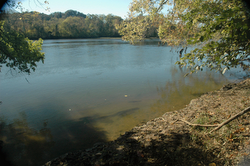
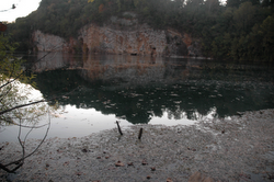
- Much of the water had evaporated (My water sample was taken from the nature quarry from Iiams Nature Park)
- The small sprigs of plant that we included in the aquarium seemed like they had grown some, or at lest had spread out across the plastic frame
- There were several microscopic organisms living amongst the plants hat were placed in the aquarium
- When observed under the microscope on the 10x objective, they appeared to be eating much the soil and then desecrating in the water
With the help of Dr. McFarland, we identifies 3 organisms:
- There was one Cyclops skeleton
-There were several Rotifers swimming amongst the soil and plant particles
-There were several Rotifers swimming amongst the soil and plant particles
-We also identified a Daphnia (Under the microscope, it appeared to have small red eyes and a flagella)
Tuesday, October 18, 2011
Water Sources:
PLANTS A and B ADDED TO MICROAQUARIUM
Letters reference the labels on the containers in the lab.
Plant A . Amblestegium sp. Moss. Collection from: Natural spring. at Carters Mill Park, Carter Mill Road, Knox Co. TN. Partial shade exposure. N36 01.168 W83 42.832. 10/9/2011
Plant B. Utricularia gibba L. Flowering plant. A carnivous plant. Original material from south shore of Spain Lake (N 35o55 12.35" W088o20' 47.00), Camp Bella Air Rd. East of Sparta Tn. in White Co. and grown in water tanks outside of greenhouse at Hesler Biology Building. The University of Tennessee. Knox Co. Knoxville TN.


WATER SOURCES
1. Tommy Schumpert Pond, Seven Islands Wildlife Refuge

1. Tommy Schumpert Pond, Seven Islands Wildlife Refuge, Kelly Lane , Knox Co. Tennessee. Partial shade exposure Sheet runoff around sink hole. N35 57.256 W83 41.503 947 ft 10/9/2011
2. French Broad River, Seven Islands Wildlife Refuge

2. French Broad River, Seven Islands Wildlife Refuge, Kelly Lane , Knox Co. Tennessee. Partial shade exposure French Broad River Water Shed N35 56.742 W83 41.628 841 ft 10/9/2011 Cladophora sp. alga in family Cladophoraceae
3. Carter Mill Park at spring source

3. Carter Mill Park at spring source, Carter Mill Road, Knox Co. Tennessee Partial shade exposure N36 01.168 W83 42.832 940 ft 10/9/2011
4. Holston River along John Sevier Hwy under I 40 Bridge

4. Holston River along John Sevier Hwy under I 40 Bridge Partial shade exposure Holston River water Shed N36 00.527 W83 49.549 823 ft 10/9/2011
5. Meads Quarry, Island Home Ave

5. Meads Quarry, Island Home Ave, Knox Co. Tennessee Partial shade exposure Rock Quarry N35 57.162 W83 51.960 880 10/9/2011
6. Dean's Woods - SpringCreek

6. Spring Creek off Woodson Dr runing throught Dean's Woods Road frontage., Knox Co. Tennessee. Partial shade exposure. Tennessee River water Shed N35 55.274 W083 56.888 848 ft 10/9/2011 Fissidens fontanus moss in stream
7. Pond at University of Tennessee Hospital. Cherokee Trail

7. Pond at University of Tennessee Hospital. Cherokee Trail. Knox Co. Knoxville TN Full sun exposure. Storm sewer sediment pond N35 56.305 W83 56.717 850 ft 10/9/2011 Chara sp. Green alga in Family Characeae.
8. Tennessee River at boat ramp across from Knoxville sewer plant

8. Tennessee River at boat ramp across from Knoxville sewer plant. Neyland Dr. Knox Co. Knoxville TN. Full sun exposure. French Broad and Holston Rivers water Sheds N35 56.722 W83 55.587 813 ft 10/9/2011
9. Pond at Sterchi Hills Greenway Trail.

9. Pond at Sterchi Hills Greenway Trail. Rife Range Rd. Knox Co. Knoxville TN Full sun exposure. Sheet runoff N36 02.687 W83 57.159 1065 ft 10/9/2011
10. Water pool below spring. Lynnhurst Cemetery

10. Water pool below spring. Lynnhurst Cemetery off of Adair Drive. Knox Co. Knoxville TN. Partial shade exposure Spring Feed Pond N36 01.357 W83 55.731 958 ft 10/9/2011
11. Fountain City Duck Pond.

11. Fountain City Duck Pond. West of Broadway at Cedar Lane. Knox Co. Knoxville TN Full sun exposure. Spring Feed Pond N36 02.087 W83 55.967 963 ft 10/9/2011
12. Water pool below spring. Fountain City Park

12. Water pool below spring. Fountain City Park west of Broadway at Hotel Ave. Knox Co. Knoxville TN. Full shade exposure Spring Feed Pond N36 02.253 W83 55.986 990 ft 10/9/2011
13. Plastic Bird Bath Pool

13. Plastic Bird Bath pool . 0.9 mile from Fountain City Pond on Fountain Rd. Knox Co. Knoxville TN Partial shade exposure N 36o02.249' W083o55.999' 1121 ft 10/9/2011
Tuesday, October 11, 2011
Setting Up The MicroAquarium
The MicroAquariums are for us to study a collection of microorganisms that we will observe over the next several weeks.
Procedure
- Obtained a MicroAquarium (Glass stand, holder, and lid).
- Using the colored dots available, as a class, we color coded the tank (Lab section, lab bench, and seat number).
- Using a pipet we extracted water from the samples provided and filled our aquariums almost to the brim with water.
- Last, we placed out aquariums on their stands and added two plants in with our water
Subscribe to:
Comments (Atom)


